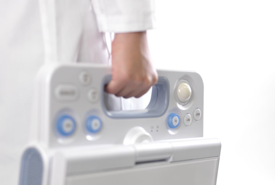
SONIMAGE HS1
From the clinic to the bedside, the SONIMAGE® HS1 delivers the superior image quality and needle visualization required for confident patient care decisions across a variety of MSK environments – orthopedics, sports medicine, physical medicine, and pain management.
The HS1, with enhanced signal penetration, increased color flow sensitivity, and improved resolution, is capable of detailed tissue differentiation, detecting structures as small as several hundred microns. In conjunction with beam-steering technology, the HS1 incorporates an advanced algorithm that utilizes both the in-plane and out-of-plane methods to improve needle visibility, especially in steep angle approaches. The resulting clarity of the needle enables increased accuracy in needle placement, making the portable system an ideal solution for pain management guided injections.
Features
ULTRAADJUST™
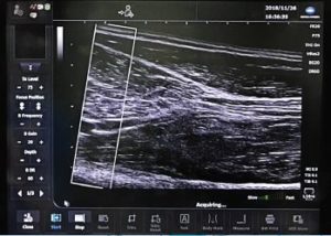
The UltraAdjust image optimization feature allows imaging parameters to be changed by simply adjusting depth. Various imaging parameters such as trapezoid setting, contrast enhancements, SI filter and focus position are automatically optimized “with the touch of the depth button”
- Improved Workflow
- MSK presets linked to anatomical structures are optimized when depth is adjusted
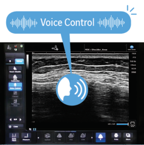
VOICE CONTROL
Voice activation of the SONIMAGE® HS1 Ultrasound System allows for handsfree operation during interventional procedures
- Simple and accurate voice activation
- Handsfree system operation
- No need to break the sterile field
- Focus more on the patient
- Enhance diagnostic efficiency
The HS1 System voice control operation feature fosters user comfort and proper operator ergonomics at the point-of-care.
PANORAMIC VIEW
Panoramic view is an imaging process that produces a panoramic image that provides both qualitative and quantitative information. Panoramic imaging stitches a series of images to give one long image in order to:
- Assess larger lesions
- Construct a cross-section image of a structure
- Show the relationship of two structures in a single image
Panoramic imaging technology widens the field of view for precise clinical diagnosis and interventional procedures.
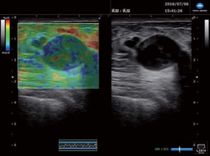
ELASTOGRAPHY
Strain Elastography is a real-time qualitative and quantitative imaging software feature that calculates and displays the relative stiffness of tissue. The image is generated from strain generated by uniform pressure of the transducer on the tissue. Use elastography to visibly assess changes in tendon and ligament elasticity.
BROAD FREQUENCY LINEAR TRANSDUCERS
Unique nanofabrication technology, combines both material and machining expertise, to produce the SONIMAGE HS1 L18-4 linear transducer. This broad frequency linear probe offers both high sensitivity as well as greater penetration, to scan both deep and superficial joints and structures.
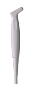
Konica Minolta releases the new HL18-4 hockey stick linear transducer for musculoskeletal (MSK) and pain management applications. The hockey stick probe reaches difficult to access areas easily with its small footprint and maneuverability. The clinician can evaluate joints in the fingers and ankle more readily with the probe’s angulation, improved control and greater contact with the anatomy. The high frequency probe provides excellent resolution in the near field for tissue differentiation and visualizes color flow with outstanding Doppler sensitivity.
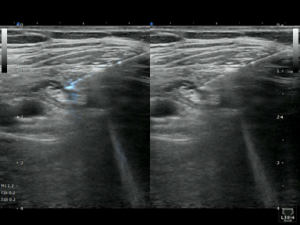
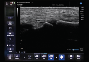
SNV™ (SIMPLE NEEDLE VISUALIZATION)
The SONIMAGE HS1 Simple Needle Visualization (SNV™) feature incorporates an advanced algorithm that utilizes both the in-plane and out-of-plane methods to improve needle visibility, especially in steep angle approaches. The resulting clarity of the needle, both tip and shaft, enables increased accuracy in needle placement, making the portable ultrasound system an ideal solution for pain management guided injections.
RHEUMATOLOGY REPORTING PACKAGE
The SONIMAGE HS1 Rheumatology report package is an efficient way to track and organize ultrasound images. Still images along with real time clips can be stored and assigned to each joint. The RA protocol is integrated and customizable. It can be operated from a foot switch allowing the user to move rapidly through the exam without taking their hands off of the probe.
REMOTE ASSIST
From the office, receive remote consultation with Remote Assist. This virtual feature can trouble shoot the system, optimize an image, provide follow up applications support, and offer educational training. And the physician and staff never have to leave the facility.
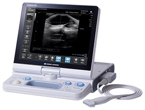
Premium Portable Ultrasound
- Imaging Performance
Excellent detail and contrast resolution for clear and homogeneous images - User friendly
8 buttons 1 touch Innovative design Intuitive user interface - 3THI
Triad Tissue Harmonic Imaging (3THI): exceptional image clarity and homogeneity - SNV
Simple Needle Visualization supports ultrasound-guided interventional procedures, with both in-plane and out-of-plane approaches - Simple clear flow
- Strain elastography
- Rheumatoid Arthritis Workflow
Unique tool supports and helps speeding up assessments of joints for Rheumatoid Arthritis